Plankton comes from the greek word planktos, meaning wanderer. It does not define a specific organism, but rather a specific life style. Plankton consist of all organisms dispersed in water that are passively driven by water currents or are subject to passive sinking process. Some of those organisms have an ability to produce oxygen and sugars using sunlight and CO2, just like terrestrial plants do. We call them phytoplankton (greek: phyton -plant ).
Phytoplankton are the wandering meadows of the ocean and an important base of the food web. Most of the phytoplankton are smaller than the width of the single human hair. They are feeding the hungry ocean, and the phytoplankton composition determines the diversity of organisms developing on the higher trophic levels like fish, birds and mammals. It is not only important for organisms living in the ocean, but it is crucial for all life on Earth. Consider the size of the oceans covering our blue planet for a moment – the amount of phytoplankton every second breath that we take the oxygen is produced by phytoplankton.
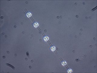
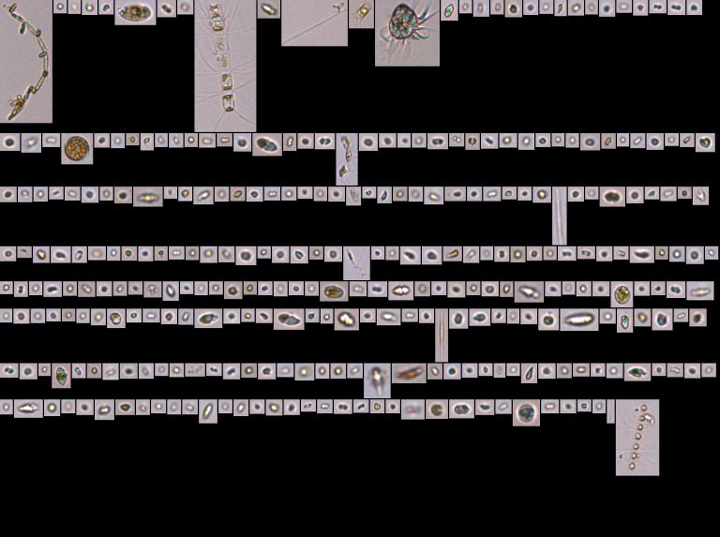
This is microscopic picture of a diatom called Leptocylindrus, a kind of phytoplankton. Phytoplankton feeds the ocean, and the phytoplankton composition determines the diversity of organisms developing on the higher trophic levels like fish, birds, and mammals. Colleen Durkin
When it comes to phytoplankton, it is not just the quantity, but also the quality that matters. You may wonder how can researchers sample something that we cannot see that exists in an environment where we cannot be in? How can we catch something that is passively driven by currents and changing all the time? How can we get the insight of the abundance of some particles that are unevenly dispersed in an ocean? Since the discovery of the microscope scientists have been trying to find answers to those questions.
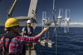
Here, on R/V Falkor we are combining traditional methods of phytoplankton analysis – such as preservation of water for later, onshore analysis under a microscope – with the new, recently developed methods of onboard image analyses. Discrete samples are taken with the Niskin bottles at the specified depth. The sampling depth is chosen according to the physical, chemical and biological characteristics of the water column that are measured by the instruments mounted on the rosette, and controlled from the science control room. Once the rosette is on deck, the samples are taken from the Niskin bottles and prepared to be either stored until they return to land, or analyzed on board. The great advantage of the onboard image analysis is that it lends an instantaneous snapshot of the phytoplankton composition and abundance, which allows you to adapt your sampling strategy and use the time spent on the ship in a better, more productive way.
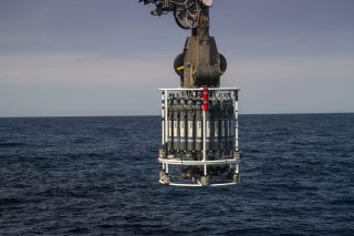
Discrete samples are taken with the Niskin bottles in each CTD cast, at specific depths. The sampling depth is chosen according to the physical, chemical, and biological characteristics of the water column. SOI/Mónika Naranjo González
The flow-through system onboard the R/V Falkor allows us for continuous sampling of the surface water for physical, chemical and biological properties, including phytoplankton composition with Imaging Flow Cytobot. This amazing instrument samples from the flow-through system every 20 minutes, and takes images of particles contained in 5 mL (100 drops) of seawater. Therefore, as we steam through the Pacific or as we stop to conduct some other measurements, we are continuously gathering high-spatial information about phytoplankton abundance and composition. The advantage of taking images vs. looking at the cells through the microscope is that you can always go back to your sample if needed. That helps us that help to have comparable results and minimizes the error of phytoplankton counting and taxonomy misidentification.
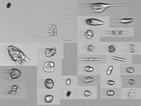
The Imaging Flow Cytobot is one of the state-of-the-art technologies used on the current expedition. As Falkor sails, a pump runs seawater through the instrument, which takes pictures of all the particles present. SOI/Ivona Cetinić
Will the modern methods of high-resolution imaging ever substitute traditional microscopy? I would say – no. While continuous sampling techniques give us amazing insight into the spatial distribution of the phytoplankton and inform best sampling strategies, classical microscopy gives us insight into the detailed morphological characteristics that are needed to be seen from multiple angles to really be understood. The phytoplankton taxonomy is under constant revision and change as the new methodologies develop. With more knowledge and deeper insight, we do find answers to existing questions, but we also encounter more questions that need to be answered. The combination of the traditional and modern methods is the best strategy to understand the secrets of these beautiful oceanic wonders.
A radiometer installed in the bow of Falkor takes color measurements from both seawater and sky. SOI/Kristen Carlson
Earth’s ocean is vast and deep, and we still need to study many things about it. To investigate and quantify biological and chemical processes, for instance, we need to determine the concentration and size of particles (living and non-living organisms) floating in the water, dissolved materials, and the diversity of organisms such as the microscopic photosynthetic phytoplankton. Their study requires both direct measurements by deploying instruments at sea or analyzing water samples, and satellite remote sensing.
Particles and dissolved organic materials scatter and absorb the sunlight that enters the ocean, which alters the ocean’s color. For instance, the first site that we sampled contained low abundances of particles including phytoplankton and dissolved organic materials, which translated to clear blue waters. Higher abundances of phytoplankton result in greener seas because of their chlorophyll pigments.
Since 1978, NASA has applied satellite remote sensing to study phytoplankton. The image depicts chlorophyll concentrations in the ocean. NASA/Norman Kuring
Since 1978, NASA has applied satellite remote sensing to study phytoplankton though its experimental Coastal Zone Color Scanner. Several other sensors followed from 1997 to the present. By 2022, NASA expects to launch the next generation ocean color satellite sensor for the Plankton, Aerosol, Cloud, and Ocean Ecosystem (PACE) which is currently being developed. This PACE sensor will provide unprecedented detail on the color spectrum and intensity of the light exiting the ocean’s surface, which will be used to infer a lot of information about our oceans, including the concentration and size of particles and dissolved organic materials, the diversity of phytoplankton, and rates of phytoplankton growth within the ocean’s sunlit surface layer.
To successfully apply the capabilities of the PACE sensor requires the development of relationships between ocean data (such as chlorophyll-a) and how it affects the color and the amount of light that will be measured by the satellite. One of our goals for participating on the Sea to Space Particle Investigation in the northeastern Pacific Ocean aboard Falkor is to collect biological, chemical and optical measurements in order to build these relationships.
To be able to do so, much of our work at sea involves development and evaluation of new methods and measurement capabilities to ensure that the data collected are of sufficient quality for application with satellite remote sensing. For example, to quantify phytoplankton growth rates, we are conducting experiments with phytoplankton and measuring the oxygen produced and carbon dioxide consumed over time.
Only satellite remote sensing can provide the comprehensive data sets across space and time needed to study the state of Earth’s vast ocean. The ocean moderates our weather, provides food, medicine, energy resources, recreation, and many other benefits. Improving our understanding of the ocean will help us better predict how it will change in the future.

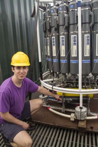
Antonio Mannino, Oceanographer, installs a Coulometer in Falkor’s wet lab to measure particle productivity in water samples collected during the expedition. SOI/Mónika Naranjo González
I always knew that one day I wanted to study the ocean, even though I grew up just north of Pittsburgh and had never seen the ocean. After graduating high school, I attended the College of Charleston in South Carolina where my plan from the start was to major in Marine Biology. I began my junior year in college with no idea what I wanted to do with this very broad degree. Then I took the required oceanography course – after that, oceanography and phytoplankton (aquatic plant life that is microscopic) were in my life permanently.
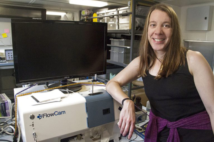
Aimee Neely, Biological Oceanographer, is studying particles using a FlowCam, an instrument that takes pictures of all the particles in the water flowing from a pump located in Falkor’s aft. SOI/Mónika Naranjo González
For an undergraduate project, I measured the response of several species of phytoplankton to different light intensities by measuring the concentration of their photosynthetic pigments, the compounds that collect light for photosynthesis. Pigments can also be used to identify specific groups of phytoplankton. During the second year of my Master’s program for marine biology, I participated in my first research cruise on a Canadian Coast Guard vessel that sailed from Dutch Harbor to Barrow, Alaska. I got my first taste of filtering, which is the collection of particles onto glass fiber pads that can be used for various analyses. Despite my initial bout of sea sickness in a near flat sea state, I was hooked.
In 2007 I pursued a research opportunity at the Bermuda Institute of Ocean Sciences where I was ship-bound once a month measuring the sulfur-based compounds made by phytoplankton that are thought to enhance cloud formation when they are outgassed to the atmosphere. For years, what I knew about phytoplankton was based on chemistry and physiology.
Samples filtered by Aimee will be processed back on land to measure different pigments in order to identify the planktonic organisms contained in it. SOI/Mónika Naranjo González
In late 2008, I left the beautiful island of Bermuda (crazy, right?) for Maryland to work at NASA Goddard Space Flight Center. Before this time, I knew nothing about satellites or ocean color remote sensing. While working at NASA I have learned that everything in the ocean – dissolved compounds, phytoplankton, and particles – absorb and scatter sunlight. Using this information about the color of the light reflecting out of the ocean, we can translate this light into information about what types of phytoplankton are in the water column.
High temporal and spatial resolution observations in the global ocean are just not feasible as we are limited by time and resources. Therefore, we make use of additional tools to fill in the gap for global and regional oceanographic observations. Satellite ocean color observations provide global ocean coverage, reaching time and space beyond our capabilities with research vessels and, therefore, may fill in the data gap where field measurements are limiting.
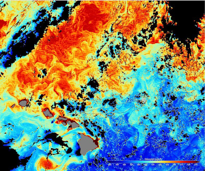
A satellite image shows Falkor’s track and the colors in ocean water. Colors indicate the amount of chlorophyll, where red is the highest and blue the lowest. SOI/Mónika Naranjo González
Ground truthing of these measurements of phytoplankton types through ocean color remote sensing is necessary but challenging. We can use phytoplankton pigments to derive a certain amount of information but the addition of microscopy is ideal, as then we can see which species are in the water. One of the newer technologies in the field is imaging flow cytometry, a technology that combines the best aspects of microscopy, flow cytometry and digital imaging.
Water is fed through the instrument at a specific magnification wherein a camera can be triggered to take a digital image of each particle or phytoplankter that passed by the field of view. Imagine how high spatial resolution of these data will help us to ground truth the phytoplankton type products that we retrieve from satellite imaging. On the RV Falkor, we have two forms of this technology to sample, not only the surface of the ocean, but also at depth. Having never spent much time in front of a microscope myself, I am learning so much from the skilled scientists around me who can look at an image and almost immediately identify to which genus and/or species the phytoplankton belongs. I hope to gain this knowledge as I learn and use this instrumentation.
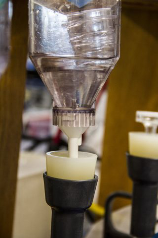
The Flow Cam is an instrument used by Aimee to identify particles in the water. Water is fed through the instrument at a specific magnification wherein a camera can be triggered to take a digital image of each particle or phytoplankter that passed by the field of view. SOI/Aimee Neeley
Melissa Omand, interdisciplinary physical oceanographer from the University of Rhode Island’s Graduate School of Oceanography, was confronted with a conflict: it was time for an upgrade to her phone, but creating more technological trash did not feel right. Plus, the camera on her older phone was fantastic. Together with her first graduate student Noah Walcutt, she worked on optimizing better battery life, as well as fabricating an underwater housing and a lighting system for her “outdated” gadget. This to her remains the best part of her job: creating and testing new instruments, as well as repurposing existing ones.
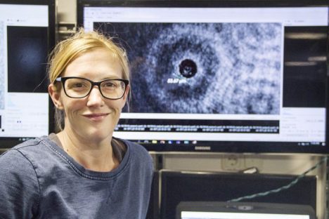
Creating and testing new instruments, as well as repurposing existing tools, are some of Melissa Omand’s favorite aspects of her job. Melissa is a Physical Oceanographer currently sailing on board R/V Falkor. SOI/ Mónika Naranjo González
Melissa and Noah are working with two different novel instruments in this cruise. The first one is a time-lapse camera developed after repurposing her previous mobile. The phone will dangle at the base of a 150 meter wire, deployed as part of the Wirewalker assembly. For three or four days, the camera snaps pictures of the base of a sediment trap which collects falling particles called marine snow. Up until now, Melissa and her colleagues Colleen Durkin and Meg Estapa have been able to identify what kind of particles fall into the traps (and at what time this happens) by analyzing the material preserved in a special gel. They have also learned that particles fall in pulses as opposed to a steady flow. However, they are still not sure about which types of marine snow sink with each pulse, and how these are connected to the phytoplankton community above. They hope the images taken by the camera will provide a new piece to complete the puzzle that is carbon storage in the ocean.
Melissa Omand deploys the sediment traps as part of the Wirewalker, while Oliver Hurdwell observes closely. SOI/ Mónika Naranjo González
A second novel instrument Melissa has brought on board is a holographic camera. Unlike traditional photography, a holographic image is obtained when a laser beam hits an object and either bounces off of it or goes through it, bending the light. More a computer than just a camera, the instrument combines diffraction data with math in order to reconstruct the light’s journey after interacting with the object (in this case, a planktonic organism). Tracing the behaviour of the light provides an enormous amount of information about the object’s characteristics in three dimensions. The result is an image that allows the experts to focus on different planes, and not in just one single depth of field.
This is interesting both because it vastly increases the volume sampled by enabling the scientists to choose where to focus on a picture that has already been taken, and in that it enables a very exciting application: Virtual Reality.
Noah Walcutt examines the holographic camera installed in the CTD rosette. The camera is able to capture around 40 000 images in a single CTD cast. SOI/ Mónika Naranjo González
Cut a single hair a hundred times and you will get something resembling the size of a few microns. That is the resolution that the holographic camera can capture in a single photo: 16 photos per second, 100 particles per hologram in average, 40 thousand holograms per dip in the ocean. The numbers begin adding up fast, so Melissa knows it is time once again to create something from scratch: their own pathway for data management, processing and analysis. This is why she began working with Ben Knorlein, a computer scientist from the Center for Computation and Visualization from Brown University.
Not only is Ben in charge of designing an efficient way to deal with all of the information yielded by the holographic camera, but he is also the mastermind behind the software that allows scientists to step into the holograms and interact with the particles in a Virtual Reality environment. This has been the first time Ben has ever been at sea. He assists researchers in the deployment and recovery of scientific instrumentation, and more importantly, he is gaining a deeper understanding of what they are looking for, becoming familiar with their thought process and expectations. All of this experience is vital for Ben to improve the software so scientists can have faster access to the information they need to extract from each holographic image.
No other ship could have given the team the opportunity to work efficiently with these images on board. Falkor’sHigh Performance Computer enables Ben to process tens of thousands of images in a single day, generating data immediately. Each day Ben sits at his working station in the Dry Lab, fine tuning parameters and settings to offer Melissa and Noah new options. Once back ashore, this cruise’s intense collaboration will have made the trio tighter than ever, and they will walk away from Falkor carrying invaluable new information, instruments and software.
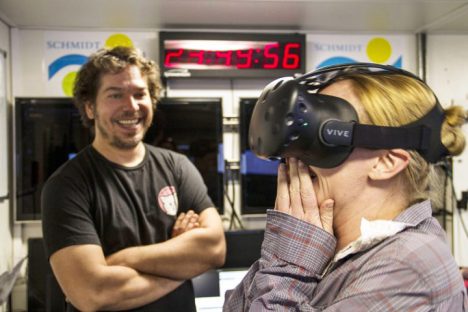
Ben Knorlein, Computer Scientist, observes Melissa Omand as she reacts to the first Virtual Reality experience created on board Falkor from holographic images of plankton suspended in the water. SOI/ Mónika Naranjo González
Trying to sleep on a trampoline while somebody is jumping on it – this is how it feels during many nights at sea as the ship zig-zags in an imaginary box around our drifting instruments in the North Pacific during winter. This is when biological activity is lowest, but clearly there is no absence of physical forces, such as waves. Clearly.
The aim of this expedition is to find phytoplankton and measure their characteristics using light detectors, cameras and microscopes. From my perspective as an oceanographer who uses satellite data to explore large scale physical forcing of biology, this is a great chance to think about the smaller-scale forcing mechanisms that supply nutrients to the phytoplankton. And I am glad to get acquainted with the optical and biological instruments and methods being used and tested here at sea.
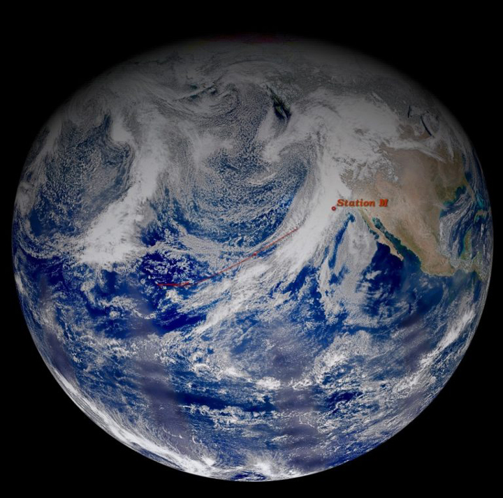
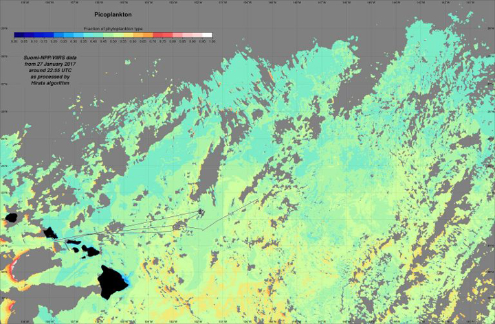
Hemispheric view by Suomi-NPP VIIRS on Feb 9, 2017 in true color. Clouds and airborne particles are white; ocean, blue. The ship’s track is shown in the red line. Station M is our last sampling site. NASA/ Norman Kuring
We began the campaign near Hawaii at the end of January in the North Pacific subtropical gyre, which has a predominate slow-moving circulation pattern that causes nutrient-depleted surface water. We experienced plenty of swell from distant storms – lab equipment had to be tied down, and chairs slid across the galley. Still, nutrient-rich deep water remained far below the well-mixed surface waters.
The water was exceptionally clear. Sunlight penetrated deeper than 150 meters (500 feet). In spite of the dearth of nutrients, our imaging systems revealed some phytoplankton! They appeared malnourished, but surprisingly diverse nonetheless.
In the absence of strong currents or other flow patterns, the Wirewalker instrument drifted westward making daily clockwise loops with the Earth’s rotation. I was excited to see its path mapped with inertial oscillations so clearly visible. Although they are always present, it is rare to see them this obviously as they are usually hidden by stronger forcing.
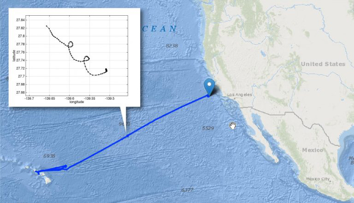
A plot of the Wirewalker’s track as it drifted freely at our second site for three days. Each point in the plot represents one hour. SOI/ Melissa Omand
The end of our sampling campaign is approximately 250km (150 miles) west of central California over a site called ‘Station M.’ This location is typically more productive, being in the California Current that brings cooler water southward from the subpolar gyre.
Additionally, we arrived between low pressure frontal systems that have been pummeling the west coast with strong winds, rain and snow over the past month. These strong weather systems cause wind-mixing at the surface of the ocean, bringing nutrients up from depth. Sampling revealed a warmer, fresher top 30 meters (100m) above cooler, nutrient-rich water.
Immediately, the instruments monitoring phytoplankton and nutrients began registering significantly higher quantities than anything we saw earlier in the expedition: even more diverse and even more abundant. Collectively, this team has gathered an amazingly rich data set of measurements and images that makes the discomfort of sleeping on a trampoline all worth it.
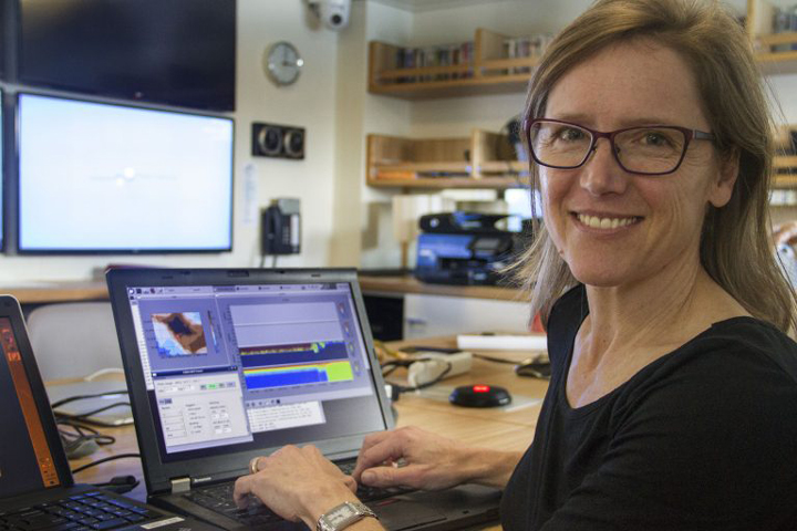
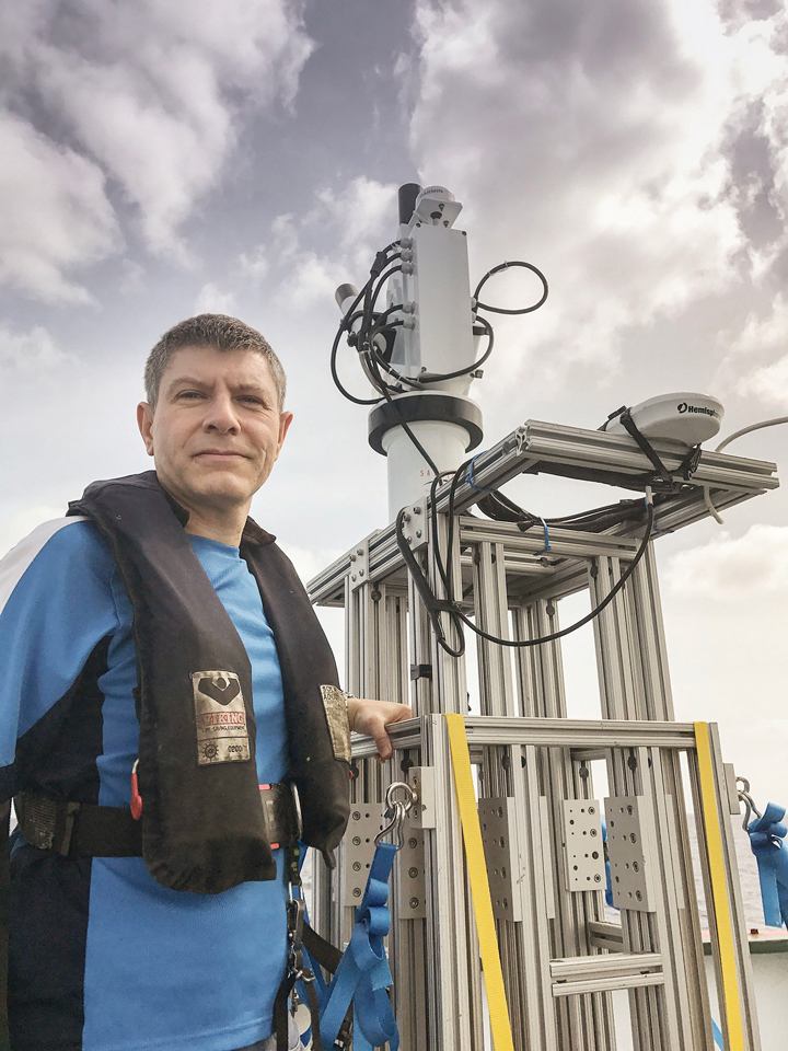
Stephanie Schollaert Uz monitors the speed and direction of water flowing under the ship with the Acoustic Doppler Current Profiler. SOI/ Monika Naranjo Gonzalez
