
I always knew that one day I wanted to study the ocean, even though I grew up just north of Pittsburgh and had never seen the ocean. After graduating high school, I attended the College of Charleston in South Carolina where my plan from the start was to major in Marine Biology. I began my junior year in college with no idea what I wanted to do with this very broad degree. Then I took the required oceanography course – after that, oceanography and phytoplankton (aquatic plant life that is microscopic) were in my life permanently.
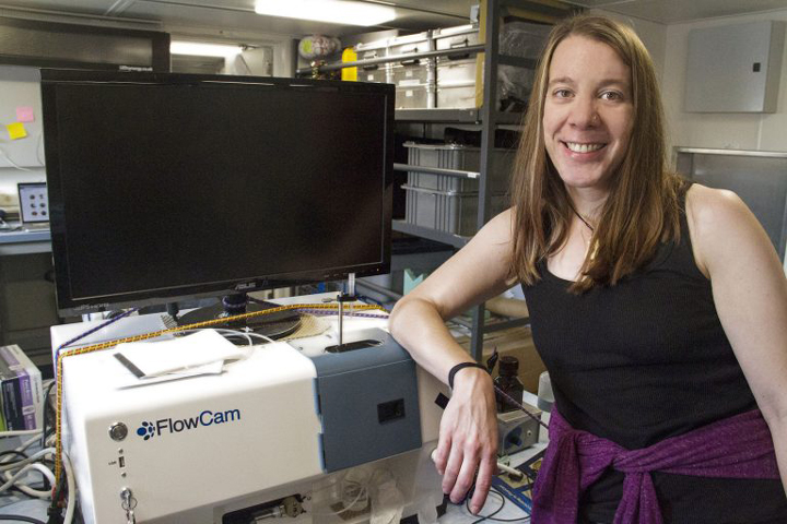
Aimee Neely, Biological Oceanographer, is studying particles using a FlowCam, an instrument that takes pictures of all the particles in the water flowing from a pump located in Falkor’s aft. SOI/Mónika Naranjo González
Phase 1: What Are Phytoplankton?
For an undergraduate project, I measured the response of several species of phytoplankton to different light intensities by measuring the concentration of their photosynthetic pigments, the compounds that collect light for photosynthesis. Pigments can also be used to identify specific groups of phytoplankton. During the second year of my Master’s program for marine biology, I participated in my first research cruise on a Canadian Coast Guard vessel that sailed from Dutch Harbor to Barrow, Alaska. I got my first taste of filtering, which is the collection of particles onto glass fiber pads that can be used for various analyses. Despite my initial bout of sea sickness in a near flat sea state, I was hooked.
In 2007 I pursued a research opportunity at the Bermuda Institute of Ocean Sciences where I was ship-bound once a month measuring the sulfur-based compounds made by phytoplankton that are thought to enhance cloud formation when they are outgassed to the atmosphere. For years, what I knew about phytoplankton was based on chemistry and physiology.
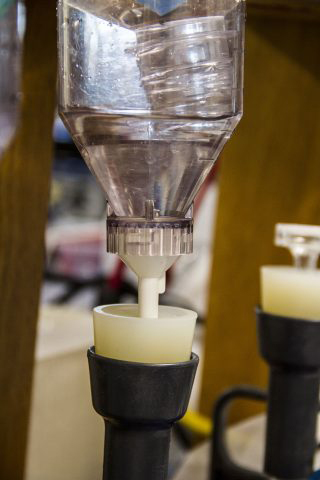
Samples filtered by Aimee will be processed back on land to measure different pigments in order to identify the planktonic organisms contained in it. SOI/Mónika Naranjo González
Phase 2: We Can Measure Phytoplankton from Space?
In late 2008, I left the beautiful island of Bermuda (crazy, right?) for Maryland to work at NASA Goddard Space Flight Center. Before this time, I knew nothing about satellites or ocean color remote sensing. While working at NASA I have learned that everything in the ocean – dissolved compounds, phytoplankton, and particles – absorb and scatter sunlight. Using this information about the color of the light reflecting out of the ocean, we can translate this light into information about what types of phytoplankton are in the water column.
High temporal and spatial resolution observations in the global ocean are just not feasible as we are limited by time and resources. Therefore, we make use of additional tools to fill in the gap for global and regional oceanographic observations. Satellite ocean color observations provide global ocean coverage, reaching time and space beyond our capabilities with research vessels and, therefore, may fill in the data gap where field measurements are limiting.
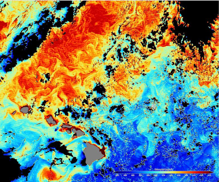
A satellite image shows Falkor’s track and the colors in ocean water. Colors indicate the amount of chlorophyll, where red is the highest and blue the lowest. SOI/Mónika Naranjo González
Phase 3: Learning How To See Phytoplankton
Ground truthing of these measurements of phytoplankton types through ocean color remote sensing is necessary but challenging. We can use phytoplankton pigments to derive a certain amount of information but the addition of microscopy is ideal, as then we can see which species are in the water. One of the newer technologies in the field is imaging flow cytometry, a technology that combines the best aspects of microscopy, flow cytometry and digital imaging.
Water is fed through the instrument at a specific magnification wherein a camera can be triggered to take a digital image of each particle or phytoplankter that passed by the field of view. Imagine how high spatial resolution of these data will help us to ground truth the phytoplankton type products that we retrieve from satellite imaging. On the RV Falkor, we have two forms of this technology to sample, not only the surface of the ocean, but also at depth. Having never spent much time in front of a microscope myself, I am learning so much from the skilled scientists around me who can look at an image and almost immediately identify to which genus and/or species the phytoplankton belongs. I hope to gain this knowledge as I learn and use this instrumentation.
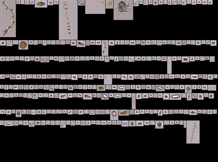
The Flow Cam is an instrument used by Aimee to identify particles in the water. Water is fed through the instrument at a specific magnification wherein a camera can be triggered to take a digital image of each particle or phytoplankter that passed by the field of view. SOI/Aimee Neeley
Tags: Carbon cycle, ocean science, phytoplankton, Sea to Space 2017